Parts of the Ear: Anatomy, Function, and Diagram
You likely know your ear does more than just hold up glasses — it’s a finely tuned machine that turns vibrations into sound and keeps you upright. This guide unpacks each part of the ear, from the visible outer folds to the tiny bones inside, and explains how they work together.
Main sections of the ear: 3 (outer, middle, inner) ·
Number of ossicles (ear bones): 3 ·
Length of the ear canal: Approximately 2.5 cm ·
Diameter of the eardrum: Approximately 8–10 mm ·
Number of cochlear hair cells per ear: About 15,000
Quick snapshot
- The ear divides into three main parts: outer, middle, inner (Jack Westin MCAT Content).
- The ossicles (malleus, incus, stapes) transmit and amplify sound vibrations (Stanford Medicine Otolaryngology).
- The cochlea converts mechanical vibrations into electrical nerve impulses (Stanford Medicine Otolaryngology).
- Exact mechanisms of age-related hearing loss (presbycusis) are still under investigation.
- Research is ongoing into hair cell regeneration in mammals.
- The three-part classification of ear anatomy has been established since the 19th century and remains the standard today (TeachMeAnatomy).
- Future treatments may target hair cell regeneration to restore hearing loss — a frontier being actively explored in labs worldwide.
The key facts below show the major numbers that define ear anatomy.
| Label | Value |
|---|---|
| Total major parts | 3 (outer, middle, inner) |
| Number of ossicles | 3 per ear |
| Length of ear canal | ~2.5 cm |
| Diameter of eardrum | ~8–10 mm |
| Smallest bone | Stapes (3 mm) |
| Hair cells in cochlea | ~15,000 per ear |
The pattern: these numbers reveal the ear’s precision engineering — each measurement is optimized for sound transmission.
What are the parts of the ear called?
Outer ear
- Pinna (auricle): The visible flap that captures sound waves and funnels them into the ear canal. It is made of elastic cartilage covered by skin (TeachMeAnatomy).
- Ear canal (external auditory meatus): A tube about 2.5 cm long that carries sound to the eardrum. Its skin contains glands that produce cerumen (earwax) for protection.
- Tympanic membrane (eardrum): A thin, cone-shaped membrane that vibrates when sound waves hit it. It marks the boundary between the outer and middle ear (Stanford Children’s Health).
Middle ear
- Tympanic membrane: Shared border with outer ear; its vibrations pass to the ossicles.
- Malleus (hammer): The first ossicle, attached to the eardrum.
- Incus (anvil): The middle ossicle, connecting malleus to stapes (Jack Westin MCAT Content).
- Stapes (stirrup): The smallest bone in the human body, about 3 mm long, that presses against the oval window of the inner ear.
- Eustachian tube: A canal that connects the middle ear to the nasopharynx, equalizing air pressure (Stanford Medicine Otolaryngology).
Inner ear
- Cochlea: A spiral, fluid-filled structure that translates vibrations into neural signals via hair cells.
- Vestibule: The central chamber of the inner ear, containing the saccule and utricle — organs that detect linear acceleration and head position.
- Semicircular canals: Three looped tubes (anterior, posterior, horizontal) filled with fluid that sense rotational head movements (Stanford Medicine Otolaryngology).
- Auditory nerve (cranial nerve VIII): Carries electrical signals from the cochlea and vestibular organs to the brainstem.
The implication: each section has a distinct job — the outer ear collects, the middle ear amplifies, and the inner ear transduces.
Each of the three ear sections has a distinct job: the outer ear collects, the middle ear amplifies, and the inner ear transduces. Damage at any stage can disrupt hearing or balance.
What are the three main parts of the middle ear?
Tympanic membrane (eardrum)
The eardrum is a thin, translucent membrane about 8–10 mm in diameter. It vibrates in response to sound waves and transfers those vibrations to the malleus. Its two layers — epithelial and fibrous — allow it to self-repair minor tears (Stanford Children’s Health).
Ossicles (malleus, incus, stapes)
- Malleus (hammer): Attached to the eardrum at its handle (manubrium); it moves when the eardrum vibrates.
- Incus (anvil): Acts as a lever between malleus and stapes.
- Stapes (stirrup): Its footplate sits in the oval window of the inner ear. The ossicles together amplify vibration force by about 1.3 times (Jack Westin MCAT Content).
Eustachian tube
This 3–4 cm tube connects the middle ear to the pharynx. It opens briefly during swallowing or yawning to equalize pressure. Dysfunction can lead to fluid buildup and otitis media (Stanford Medicine Otolaryngology).
The catch: the middle ear’s trio of bones is the only mechanical amplification stage in the auditory chain — without it, sound would lose about 30 dB of intensity before reaching the inner ear.
The middle ear’s trio of bones is the only mechanical amplification stage in the auditory chain. Without it, sound would lose about 30 dB of intensity before reaching the inner ear.
What are the parts of the inner ear called?
Cochlea
The cochlea is a snail-shaped structure about 9 mm wide at its base. Inside, the organ of Corti sits on the basilar membrane and contains approximately 15,000 hair cells per ear. These hair cells convert fluid motion into electrical signals sent via the auditory nerve (Stanford Medicine Otolaryngology).
Vestibule
Located between the cochlea and semicircular canals, the vestibule houses the saccule and utricle. These contain otolith organs that sense gravity and linear acceleration — crucial for maintaining upright posture.
Semicircular canals
Three orthogonal canals (anterior, posterior, lateral) filled with endolymph. Rotation of the head causes fluid movement that deflects hair cells in the ampulla, signaling rotational velocity to the brain (Stanford Medicine Otolaryngology).
The pattern: The inner ear packs both hearing and balance in one compact space. Damage to the cochlea impairs hearing; damage to the vestibular system causes dizziness and vertigo.
What are the three ear bones called?
Malleus (hammer)
The malleus is the largest of the three ossicles, measuring about 8–9 mm. Its handle is embedded in the tympanic membrane, and its head articulates with the incus.
Incus (anvil)
The incus sits between the malleus and stapes. It has a body and two processes; the long process connects to the stapes via the lenticular process.
Stapes (stirrup)
The stapes is the smallest bone in the human body — roughly 3 mm long and weighing about 2–4 mg. Its footplate fits into the oval window of the cochlea, transmitting vibrations into the fluid of the inner ear (Jack Westin MCAT Content).
The implication: because the stapes is so tiny, it is vulnerable to otosclerosis — abnormal bone growth that fuses the stapes to the oval window, causing conductive hearing loss.
Because the stapes is so tiny, it is vulnerable to otosclerosis — abnormal bone growth that fuses the stapes to the oval window, causing conductive hearing loss. Surgical replacement is often effective.
What is the dangling part of your ear called?
Earlobe (lobule)
The earlobe is the soft, fleshy lower part of the outer ear. Unlike the rest of the pinna, it contains no cartilage — only fibrous tissue, fat, and blood vessels. This makes it the preferred site for ear piercing and blood sampling (TeachMeAnatomy).
The implication: The earlobe’s lack of cartilage gives it flexibility and sensitivity, but also makes it prone to stretching and tearing if heavy jewelry is worn over time.
What we know and what remains uncertain
Confirmed facts
- The ear has three main parts: outer, middle, inner.
- The ossicles (malleus, incus, stapes) transmit sound vibrations.
- The cochlea converts mechanical vibrations into electrical nerve impulses.
- The semicircular canals detect rotational head movements.
What’s unclear
- Exact mechanisms of age-related hearing loss (presbycusis) are still under investigation.
- Research is ongoing into hair cell regeneration in mammals.
“The ear is composed of three main parts: the outer ear, middle ear, and inner ear. Each part plays a specific role in hearing and balance.”
— Cleveland Clinic
“The outer ear includes the pinna, ear canal, and tympanic membrane. The middle ear contains the eardrum and three tiny bones called ossicles.”
— National Institute on Deafness and Other Communication Disorders (NIDCD)
Summary
Understanding the parts of the ear is the first step toward recognizing how hearing works and what can go wrong. From the pinna’s sound-collecting curve to the stapes’s gentle push on the oval window, each structure is optimized for its job. For anyone experiencing hearing loss or balance issues, knowing which part is affected can guide conversations with an otolaryngologist — and treatment options range from earwax removal to cochlear implants to vestibular therapy. The trade-off is clear: the ear’s precision also makes it fragile, and protecting it from loud noise and infection is the best way to preserve both hearing and balance.
kenhub.com, youtube.com, youtube.com, emedicine.medscape.com
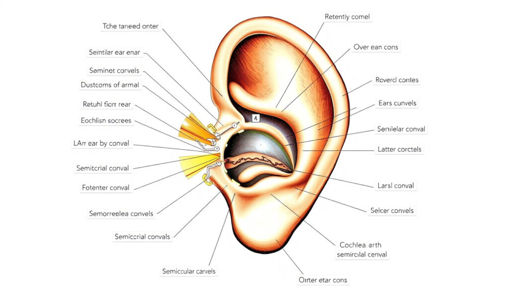
A detailed diagram of ear anatomy helps clarify how the outer, middle, and inner ear structures work together for hearing and balance.
Frequently asked questions
What is the function of the eardrum?
The eardrum (tympanic membrane) vibrates when sound waves reach it and transfers those vibrations to the ossicles of the middle ear.
How does the cochlea turn sound into electrical signals?
Fluid waves inside the cochlea cause hair cells on the basilar membrane to bend, opening ion channels that generate electrical impulses in the auditory nerve.
What are the semicircular canals responsible for?
They detect rotational movements of the head — turning, tilting, and spinning — and send that information to the brain to maintain balance.
What is the pinna made of?
The pinna (auricle) is made of elastic cartilage covered by thin skin, except the earlobe which contains no cartilage.
Why do we have earwax (cerumen)?
Cerumen protects the ear canal by trapping dust, debris, and microorganisms, and it helps keep the skin of the canal moist.
What is the Eustachian tube and what does it do?
It is a narrow passage that connects the middle ear to the back of the throat, equalizing air pressure on both sides of the eardrum.
How do the ossicles amplify sound?
The lever action of the malleus and incus, combined with the smaller area of the stapes footplate, increases the force of vibrations by about 1.3 times.
More related posts
 Tyler Perry Movies and TV Shows – Full List, Madea, Netflix Guide
 Barre Classes Near Me: Benefits, Comparisons, and More
 Eid al-Fitr 2025: Date, Meaning, and Key Practices
 Benefits of Creatine for Women: A Complete Guide to Safe Use
 12 Years a Slave – True Story, Book vs Film Facts
 Life Insurance for Seniors: Best Irish Options and Eligibility
 The Venetian Las Vegas: Suites, Costs, Perks & Fees
 49ers vs Seahawks Stats: Head-to-Head Record, Scores & History
